Human Arm Muscles Diagram / Forearm Anatomy Stock Illustrations 914 Forearm Anatomy Stock Illustrations Vectors Clipart Dreamstime - The tendons that connect the biceps muscle to the.
Human Arm Muscles Diagram / Forearm Anatomy Stock Illustrations 914 Forearm Anatomy Stock Illustrations Vectors Clipart Dreamstime - The tendons that connect the biceps muscle to the.. They are divided into two distinct compartments of the arm. Anatomynote.com found right arm muscle and tendon anatomy from plenty of anatomical pictures on the internet. The red lines show where the tendons attach the muscles to the bones. Related posts of muscles of the arm and forearm diagram piriformis muscle anatomy video. In this image, you will find frontalis, orbicularis oculi, zygomaticus, masseter, orbicularis oris, sternocleidomasteoid, deltoid, pectoralis major, biceps brachii, iliopsoas, adductor longus, gastrocnemius.
In human anatomy, the arm is the part of the upper limb between the glenohumeral joint (shoulder joint) and the elbow joint. Anatomynote.com found human arm muscle anatomy in detail from plenty of anatomical pictures on the internet. The red lines show where the tendons attach the muscles to the bones. Related posts of muscles of the arm and forearm diagram muscle anatomy chest. Many of the muscles that move the fingers and thumb originate in the forearm.
Arm Muscle Anatomy High Res Stock Images Shutterstock from image.shutterstock.com Some make broad, smooth movements, and others make small, finite movements. The upper arm is located between the shoulder joint and elbow joint. For more anatomy content please follow us and visit our website: Muscles of the shoulder and upper 12 photos of the muscles of the shoulder and upper laboratory exercise 20 muscles of the chest shoulder and upper limb, muscles of the chest shoulder and upper limb answer key, muscles of the shoulder and upper extremity, muscles of the shoulder and upper limb, overview of muscles and … arm muscles diagram The arm is one of the body's most complex and frequently used structures. Anatomists refer to the lower arm as the forearm or antebrachium. The tendons that connect the biceps muscle to the. They are divided into two distinct compartments of the arm.
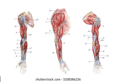
The first muscle diagram labeled 2019 above gives you an illustration of the anatomy of the arm muscle.
The first muscle diagram labeled 2019 above gives you an illustration of the anatomy of the arm muscle. Many of the muscles that move the fingers and thumb originate in the forearm. The upper arm has only one bone, the humerus, while the forearm has two, the radius and the ulna. Four muscles—the supraspinatus, infraspinatus, teres minor, and subscapularis. We hope this picture different types of muscles of arm diagram can help you study and research. For more anatomy content please follow us and visit our website: Related posts of muscles of the arm and forearm diagram muscle anatomy chest. Muscle arm… continue reading → The muscle of the arm is divided by a fascial layer separating the muscles into two osteofascial compartments: Superficial and deep anterior muscles of upper body The musculature of the forearm is complicated. The upper arm is located between the shoulder joint and elbow joint. The red lines show where the tendons attach the muscles to the bones.
The anterior and the posterior compartments of the arm. Muscle tissue diagram 12 photos of the muscle tissue diagram muscle tissue diagram, muscle tissue diagram quiz, muscle tissue structure diagram, non striated muscle tissue diagram, smooth muscle tissue diagram, human muscles, muscle tissue diagram, muscle tissue diagram quiz, muscle tissue structure diagram, non striated. Some make broad, smooth movements, and others make small, finite movements. (the lower arm is the forearm or antebrachium.) there are three muscles on the upper arm that are parallel to the long axis of the humerus, the biceps brachii, the brachialis, and the triceps brachii. The posterior (extensor) compartment contains mainly the.
Human Arm Muscular System Download Scientific Diagram from www.researchgate.net Related posts of muscles of the arm and forearm diagram muscle anatomy chest. Human anatomy and physiology lab (bsb 141) module 9: Superficial and deep anterior muscles of upper body The brachialis, biceps brachii, and brachioradialis. We'll go over all the muscles in your upper arm and forearm as well as explain. The posterior (extensor) compartment contains mainly the. The muscles of the upper arm are responsible for the flexion and extension of the forearm at the elbow joint. The image below shows the bones of the hand from the back side.
Anatomynote.com found human arm muscle anatomy in detail from plenty of anatomical pictures on the internet.
Anatomynote.com found different types of muscles of arm diagram from plenty of anatomical pictures on the internet. The muscles of the upper arm are responsible for the flexion and extension of the forearm at the elbow joint. Four muscles—the supraspinatus, infraspinatus, teres minor, and subscapularis. (the lower arm is the forearm or antebrachium.) there are three muscles on the upper arm that are parallel to the long axis of the humerus, the biceps brachii, the brachialis, and the triceps brachii. Flexion of the forearm is achieved by a group of three muscles: From the arm muscle diagram above, the muscles of the arm that can be seen easily on the surface include biceps, triceps, brachioradialis, extensor carpi radialis longus, and deltoid. The anterior (flexor) compartment contains the biceps brachii, coracobrachialis and brachialis muscles. The first muscle diagram labeled 2019 above gives you an illustration of the anatomy of the arm muscle. Human body muscle system, the muscles of the human body that work the skeletal system, that are under voluntary control, and that are concerned with movement, posture, and balance. Three of them are located in the anterior compartment — the biceps brachii, brachialis, and coracobrachialis, while the forth is located in the posterior compartment — the triceps brachii). The hand has several muscles. The arm diagram above shows the anatomy of the forearm muscles. In this image, you will find frontalis, orbicularis oculi, zygomaticus, masseter, orbicularis oris, sternocleidomasteoid, deltoid, pectoralis major, biceps brachii, iliopsoas, adductor longus, gastrocnemius.
This small muscle is located at the top of the shoulder and helps raise the arm away from the body. Labeled muscle diagram | arm muscle anatomy, muscle. In this image, you will find frontalis, orbicularis oculi, zygomaticus, masseter, orbicularis oris, sternocleidomasteoid, deltoid, pectoralis major, biceps brachii, iliopsoas, adductor longus, gastrocnemius. This diagram depicts arm muscles.human anatomy diagrams show internal organs, cells, systems, conditions, symptoms and sickness information and/or tips for healthy living. The muscle of the arm is divided by a fascial layer separating the muscles into two osteofascial compartments:
Human Arm Muscles Illustration Stock Image F011 6885 Science Photo Library from media.sciencephoto.com Some make broad, smooth movements, and others make small, finite movements. We think this is the most useful anatomy picture that you need. We hope this picture human arm muscle anatomy in detail can help you study and research. This diagram depicts arm muscles.human anatomy diagrams show internal organs, cells, systems, conditions, symptoms and sickness information and/or tips for healthy living. The arm diagram above shows the anatomy of the forearm muscles. We hope this picture right arm muscle and tendon anatomy can help you study and research. Muscle charts of the human body for your reference value these charts show the major superficial and deep muscles of the human body. We hope this picture different types of muscles of arm diagram can help you study and research.
Anatomynote.com found different types of muscles of arm diagram from plenty of anatomical pictures on the internet.
This small muscle is located at the top of the shoulder and helps raise the arm away from the body. The musculature of the forearm is complicated. This diagram depicts arm muscles.human anatomy diagrams show internal organs, cells, systems, conditions, symptoms and sickness information and/or tips for healthy living. In human anatomy, the arm is the part of the upper limb between the glenohumeral joint (shoulder joint) and the elbow joint. Human anatomy and physiology lab (bsb 141) module 9: The fascia merges with the periosteum (outer bone layer) of the humerus. Muscle anatomy upper limb 12 photos of the muscle anatomy upper limb muscle anatomy upper limb, muscle upper limb pdf, muscle upper limb table, muscles upper limb compartments, muscular anatomy of upper limb, human muscles, muscle anatomy upper limb, muscle upper limb pdf, muscle upper limb table, muscles upper limb. Your arm muscles allow you to perform hundreds of everyday movements, from making a fist to bending your thumb. Learn vocabulary, terms, and more with flashcards, games, and other study tools. The tendons that connect the biceps muscle to the. Anatomists refer to the upper arm as just the arm or the brachium. Diagram of muscles of the arm diagram muscles arm bones diagram and in the simple of. The first muscle diagram labeled 2019 above gives you an illustration of the anatomy of the arm muscle.
The image below shows the bones of the hand from the back side human muscles diagram. The (upper) arm muscles are a group of five muscles located in the region between the shoulder and elbow joints.


0 Komentar